Keratoconus is a chronic corneal pathology, in which, due to an alteration of the collagen that constitutes it causing a weakening of the cornea, thinning and deformity causing an alteration in its shape and irregular astigmatism. All this produces a progressive decrease in visual acuity, which should be avoided as much as possible with current treatments.
Typical symptoms could be: decreased vision, sensitivity to light or photophobia, rapid increase in astigmatism, and eye discomfort or irritation is very common.
This pathology is more frequent in adults and young men. It is closely related to allergic conjunctivitis, a pathology that causes the patient to rub his eyes continuously. Today we know that rubbing the eyes is one of the main risk factors for developing it.
There are several steps or phases to follow to treat keratoconus:
1.- If the keratoconus is initial, conservative treatments are chosen. The graduation of the glasses must be adjusted well, and scleral or rigid contact lenses are prescribed.
These lenses, make the deformity of the cornea is minimized when using them and thus, in many occasions, the patient can see better than with his glasses.
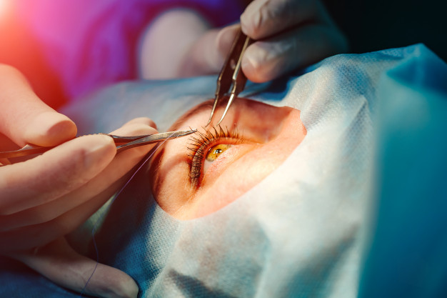
2.- Corneal cross-linking procedure (also used in the early stages of keratoconus), involves application of vitamin B2 drops followed by ultraviolet (UV) light exposure. The main objective is to increase corneal stiffness, strengthening the binding of collagen fibers.
The procedure is performed on an outpatient basis. This treatment stops or slows the progression of this pathology.
3.- Intracorneal ring segments. By placing ring segments in the peripheral area of the cornea, an attempt is made to reduce irregular astigmatism and myopia originated, trying to give the cornea a morphology as normal as possible and achieve an increase in the patient’s vision.
This treatment can be combined with Cross-linking. The placement of these implants is quite safe today, it is done with Femtosecond laser (replacing the manual form that was used years ago). The eye surgeon prepares the laser with the exact depth and location, where it is necessary to implant the ring segments to the patient’s cornea, and with the laser, intracorneal tunnels are carved where these ring segments will be housed.
Today it is possible to eliminate more diopters with the use of associated pre-crystalline intraocular lenses. So that the patient improves vision and at the same time recovers a more anatomical shape of the cornea.
If another procedure is necessary, such as corneal transplantation, which requires the cornea to being in a virgin state, intracorneal ring segments can be removed.
4.-When this pathology is in more advanced phases, and no other procedure has been effective with these patients, the Ophthalmologist specialized in cornea, performs a corneal transplant. Currently, techniques that partially replace the thickness of the cornea are used instead of performing penetrating keratoplasties (or, of the entire corneal thickness), as years ago, in this way the patient will have a faster visual recovery, and the astigmatism caused by the technique becomes less.
Gloria Carretero León Eye Clinic and Surgery
Ophthalmologist and Ocular Surgeon Marbella
Recent Comments