The pars plana vitrectomy is one of the most important employed techniques in vitreoretinal surgey within the multiple techniques in ophthalmology, which are performed to treat the back of the eye or vitreous chamber.
This surgery is performed by retina specialists, and is to remove the vitreous (or inner gel of the eye), it is because it fills with blood due to a vitreous hemorrhage. It may also because it is necessary to do it to treat the retina in cases such as: retinal detachment or macular pathologies such as the macular hole or the epiretinal membrane.
A wide variety of pathologies that require a vitrectomy, which we explain below:
-Diabetic retinopathy. It is caused by blood in the vitreous or hemovitreous chamber that cannot be reabsorbed, and also due to long-standing diabetes, cases of tractional retinal detachment.
-Rhegmatogenous detachments.
-Macula pathologies (macular hole, vitreoretinal traction or epiretinal membrane).
-Complications as a result of other surgeries, for example cataract surgery, in which there is a posterior capsular rupture with crystalline remains to vitreous.
– Endophthalmitis treatment, whether infectious or inflammatory. In this pathology, when the infection or inflammation is cooled, a large number of vitreous veils are observed and cause blurry vision even more.
– Retinal venous obstructions are another cause of vitreous bleeding.
– Eye trauma.
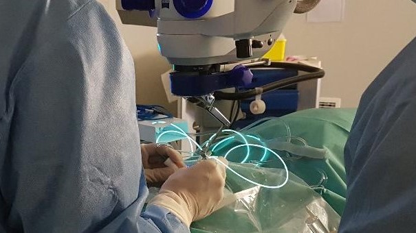
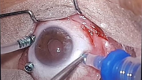
This surgery is usually performed as a major outpatient surgery with local anesthesia and sedation. The surgeon makes three small microincisions and introduces three very thin trocars serving as guides to the vitreous gel, Through the trocars all the necessary elements used in this technique are introduced, which are: an irrigation that keeps the eye at stable pressure; light to see through the microscope, and vitreotome to cut the vitreous, as well as scissors, tweezers and all the necessary instruments.
Once the surgery has been performed, it is sometimes necessary to leave the eye filled with expanding gas, this is done to achieve the quickest reapplication of the retina. The gas is reabsorbed over time, and in the most difficult cases, it is necessary to leave a substance of higher density such as silicone oil. This oil is not reabsorbent and must be removed from the eye in the long term when the ophthalmologist deems it appropriate.
The day after surgery, a checkup is necessary to remove the eye dressing and control eye inflammation and eye pressure.
After surgery, the treatment consists of putting drops into the eye and they are antibiotic and anti-inflammatory eye drops and this treatment is maintained for approximately the first month. After the first week, a practically normal life begins avoiding severe physical exercise.
Visual recovery is slow and to appreciate an improvement in vision, it is necessary to wait for the gas to be reabsorbed or for the extraction of the silicone oil.
Recent Comments